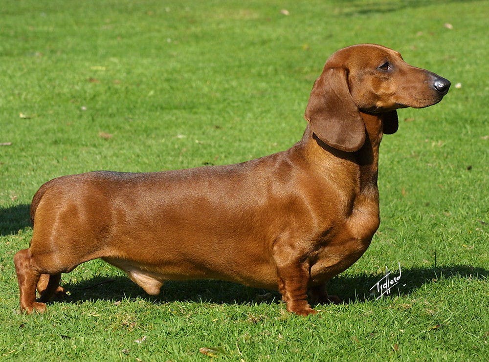
Patches, a nine-year-old dachshund, is now cancer-free thanks to a group of researchers.
Veterinary surgical oncologist Michelle Oblak from the University of Guelph’s Ontario Veterinary College and Cornell University’s small-animal surgeon Galina Hayes were the researchers who accomplished this veterinary first.
They worked in collaboration with medical technological company Adeiss to design and make a 3D-printed plate, which was intended to replace a large portion of the dog’s skull.
Patches suffered from the presence of a large tumor dangerously pressing against her brain and eye socket. The tumor was so large that it had started growing into her skull, causing a significant portion of her skull to need to be removed.
Through the use of rapid prototyping and 3D-printed implants for reconstruction, Oblak was able to map out the tumor and practice the surgery on a 3D-printed model of Patches’ brain and tumor prior to performing it.
“I was able to do the surgery before I even walked into the operating room,” Oblak said in a statement from the university.
Additionally, Oblak pointed out that 3D printing allowed for the implant to be designed specifically for Patches. In the past, the implant would have been made in a more generic way: the titanium mesh would first be molded into a general model and then modified to the patient.
“[3D printing] shifts the focus from an implant that has been designed for common use that requires modification to a patient, to a patient-specific implant that has been designed directly for them,” Oblak said, according to CNN.
This is a topic that the Carnegie Center for Surgical Innovation is actively exploring.
The lab is located in the heart of the Hopkins Hospital and was formed as a collaboration between the Department of Biomedical Engineering and Department of Neurosurgery.
The 3D Printing Facility has the ability to create material and digital models of medical conditions, patient-specific prostheses, surgical guides and functional device prototypes. This facility was created with the aim to help medical practitioners, researchers and students create sophisticated models of their ideas.
One of their affiliated labs, the I-STAR Lab, is currently conducting research in a variety of areas, which include: imaging physics, 3D image reconstruction, novel imaging systems, and image-guided interventions and diagnostic radiology. I-STAR is headed in part by Jeffrey Siewerdsen.
The Hopkins School of Medicine’s Department of Art as Applied to Medicine also works in collaboration with the Carnegie Center for Surgical Innovation.
The Department of Art as Applied to Medicine attempts to provide clear, accurate visuals to teach new surgical procedures, explain newly discovered molecular mechanisms, describe how a medical device works and depict a disease pathway.
Their work is intended to bridge the gap between medical and healthcare communication by creating 3D models that include teaching models of anatomic, cellular and molecular structures, surgical pre-planning models, engineering prototypes, custom printed lab devices, and patient education models.
In addition to using 3D printing to create implants and practice procedures, the technology is increasingly being used to educate patients.
“Patients gain a better understanding of their condition when they hold a model of their heart or brain tumor,” Siewerdsen said in an interview with the Hopkins Department of Biomedical Engineering. “It gives an entirely different level of understanding than just looking at an MRI scan.”
Dedication to researching the application of 3D printing to medicine at universities across the world has allowed for surgeries such as Patches’ to be successfully performed.
Patches has now been cancer-free for six months, said her owner, Danielle Dymeck. Although Patches slipped a disk in her back shortly after the procedure, Dymeck remains positive.
“She was just ready to be a dog again,” Dymeck said in an interview with CNN. “Cancer research like they are doing is very important to both humans and animals.”


