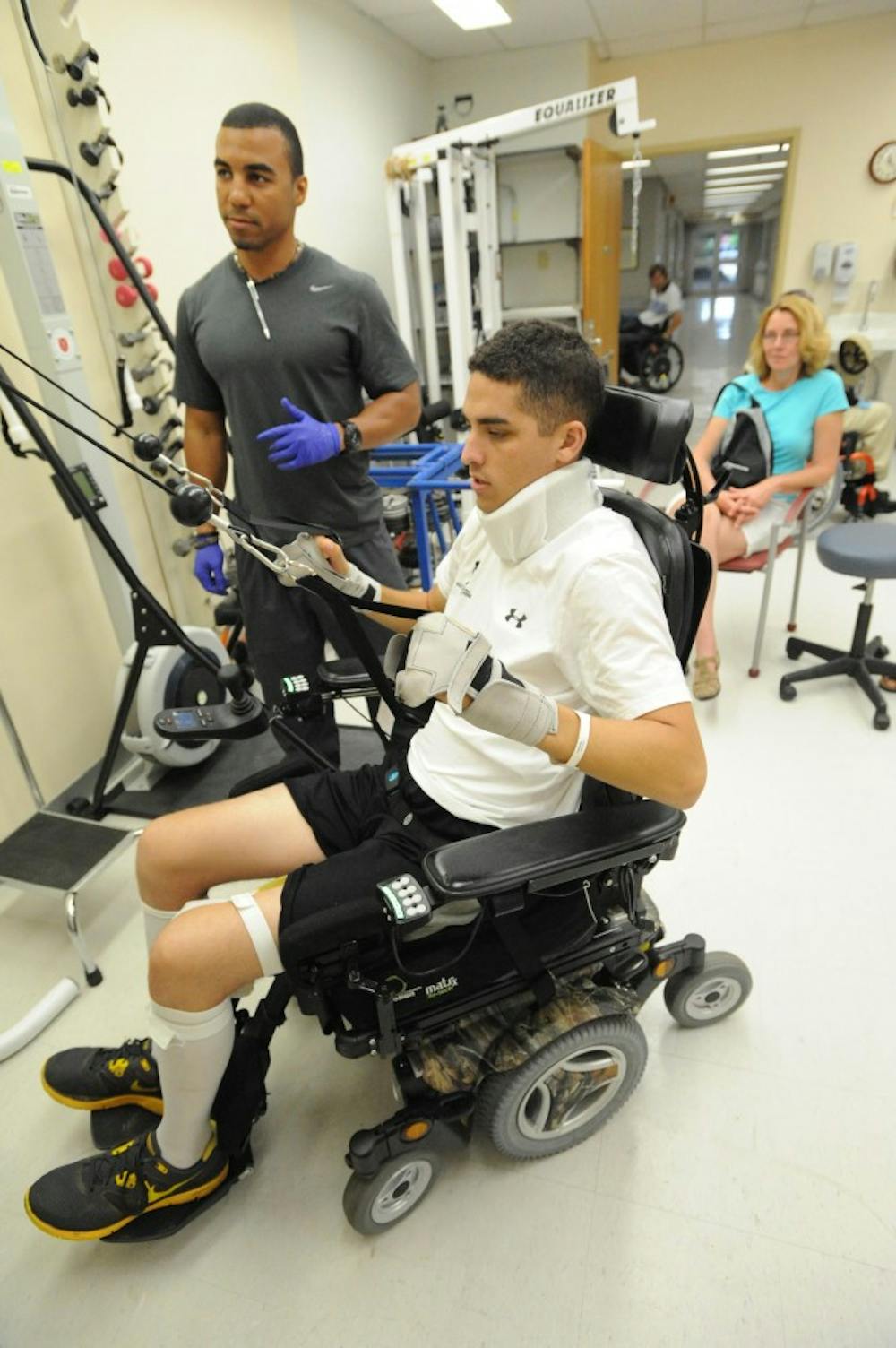
Spinal cord injuries are among the most alarming and unpredictable of injuries, carrying the possibility of paralysis. Compounding this fact, since severed nerve fibers in the central nervous system, which comprises the brain and spinal cord, do not regenerate, the paralysis has the potential to be permanent.
Like a cut wire, severed nerve fibers mean that signals from the brain cannot reach areas of the body that extend beyond the site of injury.
Recently, however, scientists from the Ecole polytechnique federale de Lausanne (EPFL) in Switzerland, the University of California, Los Angeles (UCLA) and Harvard University have begun working on a way to regenerate these nerve fibers.
Their method involves three parts, all of which are crucial for the regeneration to work.
“Our aim was to replicate, in adults, the conditions that encourage the growth of nerve fibers during development,” EPFL researcher Gregoire Courtine said in a press release.
As stated in the paper, published in Nature, the scientists showed that neuron intrinsic growth capacity, growth-supportive substrate and chemoattraction are necessary and sufficient to stimulate regrowth of nerve fibers, also known as axons.
To demonstrate the first part, the researchers used viral vectors to inhibit phosphatase and tensin homologue (PTEN), a protein that negatively regulates pathways involved in axon elongation. They also expressed proteins, including insulin-like growth factor 1 (IGF1) which have been shown to increase axon number, size and density.
To create a favorable environment for the axons to grow, the scientists delivered proteins that would help increase substrates such as laminin, which promotes axon growth.
To chemoattract axons, the researchers deposited a protein called glial-derived growth factor (GDNF), which promotes survival of axons and is able to prevent the apoptosis, or programmed cell death, of axons that have been cut.
They also showed that depositing the protein into the neural tissue below the injury site promoted axon growth past the injury.
The scientists conducted experiments in both mice and rats, injecting the three sets of molecules at different times. The three-pronged combination stimulates the axons genetically, creating a good environment and laying a trail for the nerve fibers to improve electrical conductivity in the spinal cords of the animals.
Following treatment, the animals’ spinal cords were stimulated above the injury with an electrical current, and the researchers found that axons which had been regenerated conducted 20 percent of the normal electrical signals below the injury, whereas untreated animals had no transmission.
The animals, however, were not yet able to walk.
Michael Sofroniew, a professor at UCLA and a corresponding author of the study, theorized in a statement to United Press International that the newly regrown axons were acting like axons grown during normal development and would need to be retrained.
There are some issues that need to be addressed before the therapy has the possibility to be applied in humans. Significantly, the first part of the treatment, involving the proteins that influenced the axons genetically, was administered to the animals two weeks before injury was induced, rather than after.
While further research will need to be done, the study offers a promising framework that can inform how scientists should approach stimulating axon growth and regeneration after injury.


