Imagine yourself sitting comfortably in a car, gazing through the window at the oak trees gradually coming into sight and then vanishing quickly in a blur as the wheels continue in its steady, rolling motion. A flash of light blinds your eyes momentarily, fear immediately disperses across your entire body. The airbag in front bursts out shortly, and a huge mass crashes into your door.
The next day you wake up feeling numb chest down with each limb feeling 100 pounds heavier. You see your fingers intact but motionless. Frustrated, you realize only your neck and head moves while the rest of your body is strapped down by invisible chains.
According to the Mayo Clinic, “auto and motorcycle accidents are the leading cause of spinal injuries, accounting for more than 35 percent new spinal cord injuries each year.”
Paralysis due to spinal cord injury inhibits one’s ability to perform basic motor functions, thereby reducing quality of life.
Although paralyzed individuals can have perfectly functional brains and peripheral nerves, electrical signals between the two cannot flow due to spinal cord damage. They are often overwhelmed by various emotions such as embarrassment, helplessness or fearfulness. Therefore, restoring basic movements like grasping or walking is critical to them as a means of returning to a normal life.
Researchers from the University of Utah and Oregon State University joined forces to bring new hope for paralyzed patients in a study recently published in the journal Frontiers in Neuroscience.
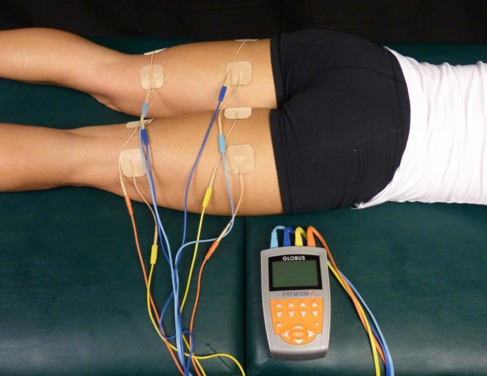
Functional neuromuscular stimulation (FNS) is a popular method that has been researched intensively by scientists who are working to restore basic motor functions in paralyzed individuals.
However, FNS devices are “limited by rapid muscle fatigue due to high stimulation frequencies and inverse recruitment of fast-fatiguing fibers, as well as and poorly evoked movement kinematics,” according to the paper published by the research group.
The researchers conducted the experiment by using a proportional-integral-velocity (PIV) controller, which sends electrical pulses at a fixed rate using an error-feedback loop, and applying the method of asynchronous intrafascicular multielectrode stimulation (aIFMS) on anesthetized cats, which is a commonly used model for human paralysis.
aIFMS allows the smooth, fatigue-resistant muscles to work by stimulating multiple independent peripheral nerve motor axons at a “lower stimulation amplitude and low per-electrode frequency” asynchronously. This technique is better able to imitate naturally occurring biochemical processes in muscles compared to the FNS technique.
Additionally, the development of the Utah Slanted Electrode Array (USEA) allowed the researchers to selectively activate the specific motor axons of interest. During the setup of this experiment, a 100-electrode stimulation device was implanted in the left sciatic nerve of the cat. Their results were successful in evoking steps in joint position, according to the researchers.
“Early versions of this technology could be used to help the person get up, use a walker and make a few steps. Even those kinds of things would have an enormous impact on someone’s life, and of course we’d like people to do more,” V John Mathews, a professor of Electrical Engineering and Computer Science at Oregon State University who contributed to this experiment, said in a press release. “My hope is in five or 10 years there will be at least elemental versions of this for paralyzed persons.”


