This week the Nobel Assembly at Karolinska Institutet awarded the Nobel Prize in Physiology or Medicine, and The Royal Swedish Academy of Sciences awarded the Physics and Chemistry Prizes. The science prizes recognized achievements from nine scientists over the past 50 years.
Physiology or Medicine
What was the discovery? Neurons called “place cells” in the hippocampus region of the brain create a map of your surroundings to provide a reference for spatial navigation (O’Keefe). “Grid cells” in the entorhinal cortex form a grid that encodes the space, allowing us to orient ourselves in a room (Moser and Moser).
How did they do it? The laureates measured the activity of individual neurons in rats to observe the firing patterns as the animals moved around an enclosed space.
What is the significance? The hippocampus is the center of memory in the brain, so understanding how we remember our surroundings will help us understand diseases like Alzheimer’s and dementia that affect these cognitives processes.
Physics
What was the discovery? The laureates figured out how to make blue light-emitting diodes (LEDs). For thirty years, we only had red and green diodes, but now, with blue as well, we have been able to create white LED lamps.
How did they do it? To create an LED, you need to grow crystals that will emit the light you want, but the semiconductor in the LED damages gallium nitride (the crystals used for blue light). The laureates discovered a method of growing gallium nitride to be of high enough quality that it can be used in an LED.
What is the significance? LED lamps are far more energy-efficient and environmentally friendly than incandescent or fluorescent lamps because they convert electricity directly to light without losing any of it to heat during the process.
Chemistry
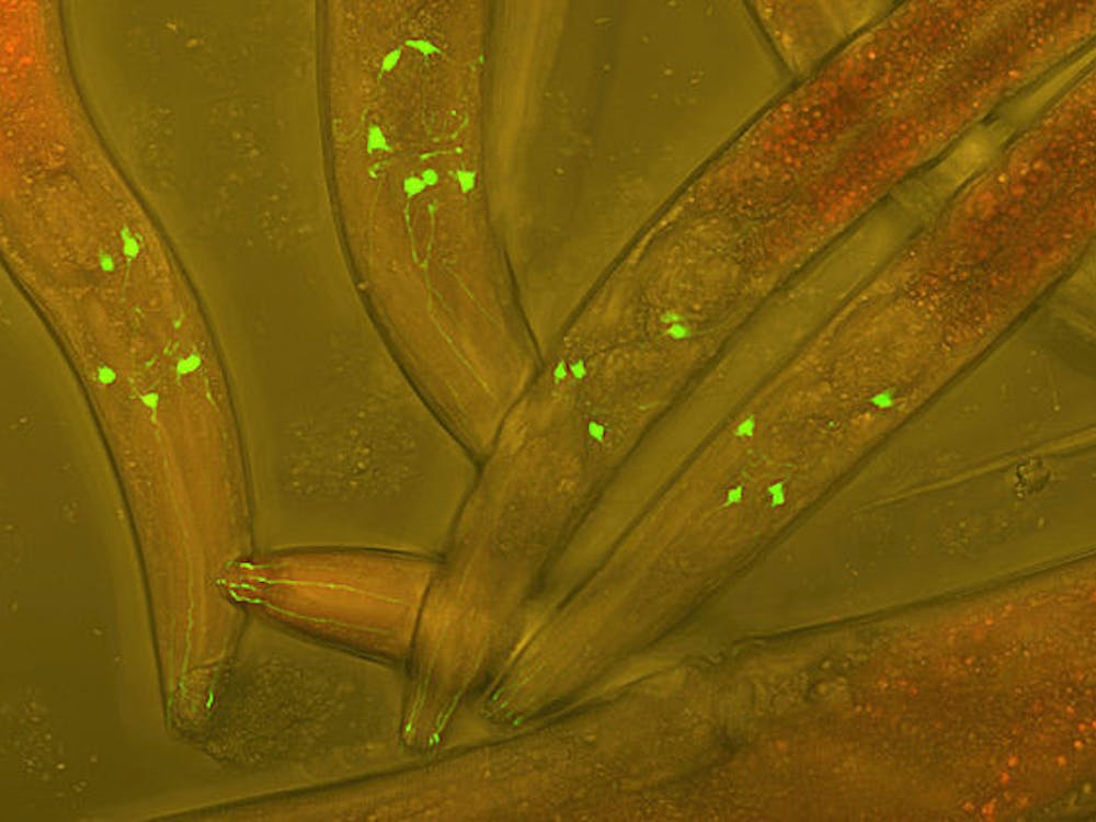
What was the discovery? The laureates contributed to current methods of observing very small subcellular structures under a microscope through illumination of individual fluorescent molecules.
How did they do it? Stimulated emission depletion (STED) microscopy excites the sample with light but blocks it out everywhere except in a very tiny region. Lots of small images put together then make up a picture of the whole sample (Hell). Single-molecule microscopy activates different fluorescent molecules at different times and then superimposes the images (Moerner and Betzig, separately).
What is the significance? For decades, we thought that there was a limit to the smallest things we could see under a light microscope — 200nm, half the wavelength of visible light. With STED and single-molecule microscopy, we can observe the proteins and organelles of a cell with higher resolution that was ever thought physically possible.


